[ad_1]
Researchers from Charité — Universitätsmedizin Berlin have been able to demonstrate how, on a molecular level, a specific protein allows light signals to be converted into cellular information. Their findings have broadened our understanding of the way how plants and bacteria adapt to changes in light conditions, which regulate essential processes, such as photosynthesis. Their research has been published in the current issue of Nature Communications.
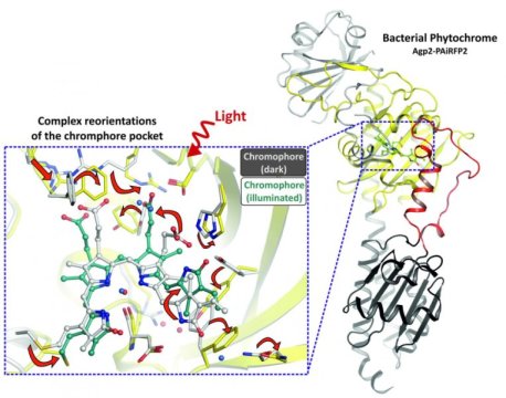
Phytochromes are proteins, which are responsible for converting light into cellular information. Found in plants, fungi and bacteria, these photoreceptors use light to regulate fundamental physiological processes. Phytochromes contain a light-sensitive tetrapyrrole molecule known as chromophore, which changes its form when exposed to light of a very specific wavelength. The protein detects these changes and implements further structural rearrangements. Activation and deactivation pathways triggered in response to light result in phytochrome undergoing a complex process of structural transformation.
Researchers from Charité’s Institute of Medical Physics and Biophysics were able to shed light on the structural transformations taking place. The researchers used X-ray crystallography to determine the 3D structure of a dark-adapted phytochrome photoreceptor and went on to compare this structure with its light-adapted state. To do this, the researchers started by creating a crystalline form of the protein, which they then irradiated with X-rays. By protein structural analysis the researchers were able to calculate the position of atoms inside the molecule. Results of their work show the contribution of individual amino acids in the light-induced activation and deactivation of these proteins. “Our research has delivered fundamental structural data, which will enhance our understanding of the way how environmental signals are transmitted into an organism. These are important insights, particularly if we hope to be in a position to use photoreceptors for future clinical applications,” explains the study’s lead researcher, Dr. Patrick Scheerer.
One potential application would be in the field of oncology, where photoreceptors might be used to visualize cancerous tissues. This particular application would be based on their ability to absorb and emit light in the red and near-infrared regions of the visible spectrum. Given that near-infrared light has a greater depth of penetration in human tissues, phytochromes could be used to visualize deeper-lying tissue cells in a non-invasive and side effect-free manner. Photoreceptors could also prove suitable as light-controlled tools, which could be used to treat genetic diseases at the molecular level. In order to explore these potential applications further, Dr. Scheerer and his team hope to use future research studies to gain a better understanding of phytochrome fluorescence (another property of these photoreceptors), as well as exploring other aspects of their structural transformation.
Story Source:
Materials provided by Charité – Universitätsmedizin Berlin. Note: Content may be edited for style and length.
[ad_2]


