[ad_1]
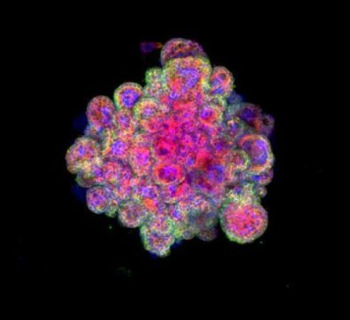
Researchers say that new ‘mini-placentas’ — a cellular model of the early stages of the placenta — could provide a window into early pregnancy and help transform our understanding of reproductive disorders. Details of this new research are published today in the journal Nature.
Many pregnancies fail because the embryo does not implant correctly into the lining of the womb (uterus) and fails to form a placental attachment to the mother. Yet, because of the complexities of studying this early period of our development, very little is understood about what is happening normally and what can go wrong. Animals are too dissimilar to humans to provide a good model of placental development and implantation.
“The placenta is absolutely essential for supporting the baby as it grows inside the mother,” says Dr Margherita Turco, the study’s first author, from the Departments of Pathology and Physiology, Development and Neuroscience at the University of Cambridge. “When it doesn’t function properly, it can result in serious problems, from pre-eclampsia to miscarriage, with immediate and lifelong consequences for both mother and child. But our knowledge of this important organ is very limited because of a lack of good experimental models.”
Efforts to grow human placental cells started over 30 years ago in the Pathology department where Professors Ashley Moffett and Charlie Loke were studying cellular events in the first few weeks of pregnancy. With their chief technician, Lucy Gardner, they found ways to isolate and characterise placental trophoblast cells. These techniques, combined with the organoid culture system, enabled the generation of miniature functional models of the early placenta — or ‘mini-placentas’.
In the past few years, a new field of research has blossomed that uses these organoids — often referred to as ‘mini-organs’ — enabling insights into human biology and disease. At the University of Cambridge, one of the world leaders in organoid research, scientists are using organoid cultures to grow everything from ‘mini-brains’ to ‘mini-livers’ to ‘mini-lungs’.
In a study funded by Wellcome and the Centre for Trophoblast Research, the Cambridge team was able to grow organoids using cells from villi — tiny frond-like structures — taken from placental tissue. These trophoblast organoids are able to survive long-term, are genetically stable and organise into villous-like structures that secrete essential proteins and hormones that would affect the mother’s metabolism during the pregnancy. Further analysis showed that the organoids closely resemble normal first-trimester placentas. In fact, the organoids so closely model the early placenta that they are able to record a positive response on an over-the-counter pregnancy test.
Professor Graham Burton, a co-author and Director of the Centre for Trophoblast Research, which last year celebrated its tenth anniversary, says: “These ‘mini-placentas’ build on decades of research and we believe they will transform work in this field. They will play an important role in helping us investigate events that happen during the earliest stages of pregnancy and yet have profound consequences for the life-long health of the mother and her offspring. The placenta supplies all the oxygen and nutrients essential for growth of the fetus, and if it fails to develop properly the pregnancy can sadly end with a low birthweight baby or even a stillbirth.”
In addition, the organoids may shed light on other mysteries surrounding the relationships between the placenta, the uterus and the fetus: why, for example, is the placenta able to prevent some infections passing from the mother’s blood to the fetus while others, such as Zika virus, are able to pass through this barrier? The organoids may also be used for screening the safety of drugs to be used in early pregnancy, to understand how chromosomal abnormalities may perturb normal development, and possibly even provide stem cell therapies for failing pregnancies.
Last year, the same team supported by Cambridge’s Centre for Trophoblast Research reported growing miniature functional models of the uterine lining.
“Now that we’ve developed organoid models of both sides of the interface — maternal tissue and placental tissue — we can start to look at how these two sides talk to each other,” adds Professor Ashley Moffett.
Professor Moffett also co-directed a recent study published in Nature that used genomics and bioinformatics approaches to map over 70,000 single cells at the junction of the uterus and placenta. This study revealed how the cells talk to each other to modify the immune response and enable the pregnancy, presenting new and unexpected cell states in the uterus and placenta, and showing which genes are switched on in each cell.
[ad_2]


