[ad_1]
While amyloid plaques have long been closely associated with mechanisms driving Alzheimer’s disease, visualizing how amyloid proteins assemble continues to prove difficult. The nanometer-sized amyloid fibrils are only a fraction of the size that the best light microscopes are able to resolve. New work repurposing one of the oldest known reagents for amyloid looks to help provide a clearer picture of how fibrils come together.
A team of researchers from Washington University in St. Louis, U.S.A., and University College London in the U.K., has demonstrated a novel approach for nanoscale imaging of amyloid structures without chemically altering them. Using Thioflavin T (ThT), a dye known for nearly a century to fluoresce when in contact with amyloid fibrils, the new method allows researchers to visualize proteins associated with amyloid plaques, called A?42 and A?40, more precisely than ever.
Kevin Spehar, a lead co-author from the team, will describe their work in an oral presentation, titled “Long-Term Super-Resolution Imaging of Amyloid Structures Using Transient Binding of Thioflavin T,” at the OSA Biophotonics Congress: Optics in the Life Sciences meeting in Tucson, Ariz., U.S.A., 14-17 April 2019.
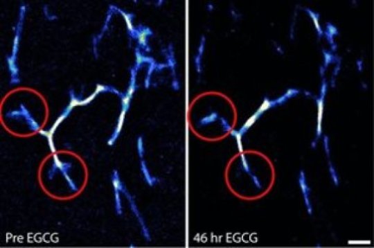
In addition to producing images of amyloid aggregates with nanoscale resolution, the group’s technique lets researchers take snapshots of how fibrils build up and react to their environment. Testing their approach, the team was able to see directly for the first time an anti-amyloid drug at work.
“When it comes to amyloid, we use words like ‘monomer’ and ‘oligomer’ and ‘fibril,’ but those words only really describe what we’ve been able to see before,” said paper co-author Dr. Matthew Lew. “Those words are completely inadequate for precisely describing the complex, varying assemblies of these molecules.”
While attacking amyloid assembly methods stands out as a leading proposed therapy for Alzheimer’s disease, Dr. Jan Bieschke, another co-author of the paper, said that studying amyloid aggregates presents unique challenges for researchers.
Immunofluorescent techniques, which are employed in many other areas of biology and use antibodies to label biomolecules, fall short because they would disrupt amyloid’s tendency to aggregate, making it impossible to accurately study the mechanism that drives Alzheimer’s.
Cryoelectron microscopy offers superior resolution but can only provide a single, static snapshot of an amyloid sample.
“Imaging amyloid dynamics for extended times is crucial if we want to understand the way a drug affects amyloid aggregation or how it disassembles an amyloid fiber,” Bieschke said.
To tackle these issues, the team turned to the long-established fluorophore, ThT, which avoids modifying amyloid by not covalently binding to it in the first place. Instead, each ThT molecule fluoresces for about 15 milliseconds while in contact with amyloid.
The result, Lew said, is that ThT’s role in imaging shifts from a simple fluorophore to a molecular sensor for amyloid.
“This is literally using a one- to two-nanometer molecule as a sensor,” he said. “I think this concept has a lot of potential to be generalized for biomedical and chemical imaging applications.”
The imaging let the team watch how A?42 fibrils remodeled and dissolved with the introduction of epi-gallocatechin gallate, a model anti-aggregation drug Bieschke and colleagues discovered.
“Most fluorescence microscopy techniques, especially when aiming for nanometer resolution, require careful fine-tuning of reagents and conditions,” Bieschke said. “Our approach removed much of that complexity. At the same time, it can be combined with traditional antibody-based approaches for multiplexed imaging.”
Bieschke hopes to improve the technique to be able to see the way amyloid structures spread in Alzheimer’s and related diseases. Lew said he sees many future uses for using molecules like ThT as molecular sensors, ranging from research in Parkinson’s disease to diabetes to materials science.
[ad_2]


